


This article was published on Arbona Health Hub Volume 1 Issue 1 (ISSN: 3065-5544).
When hearing the word nuclear, what comes to mind? Usually This feels like it belongs in your personal statement and not in this section. You can talk about how taking care of your dad reinforced your desire to study medicine, etc., one thinks about nuclear weapons or nuclear power plants, not an everyday diagnostic tool in healthcare. Nuclear medicine doesn’t share many similarities with these, but the main thing they all have in common is radiation. Nuclear medicine is a specialty that requires completion of a medical degree (MD or DO) and then 4 years of residency training. It shares similarities with Diagnostic Radiology. However, the main difference is that in nuclear medicine, radioactive material, called radioactive tracers, is used for diagnostic purposes, to monitor the health of organs/tissue, and to target and destroy diseased or damaged organs/tissue.
What are radioactive tracers?
Radioactive tracers are carrier molecules that are closely bound to a radioactive atom. The carrier molecules can differ depending on the objective of the scan. Most nuclear medicine diagnostic procedures include administering a radioactive tracer to a patient via inhalation, oral consumption, or injection into an organ. The nuclear medicine physician will choose the most accurate tracer for a given patient, tailoring it so that each patient receives an individualized assessment. The tracer used determines whether the patient will have a Single Photon Emission Computed Tomography (SPECT) or Positron Emission Tomography (PET) scan.
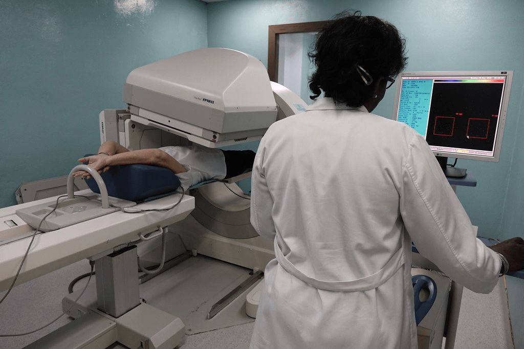
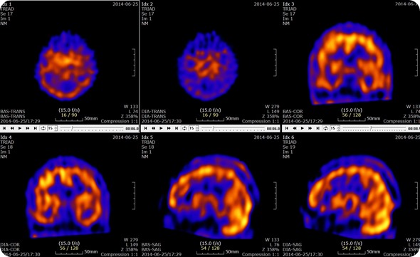
What is a SPECT scan?
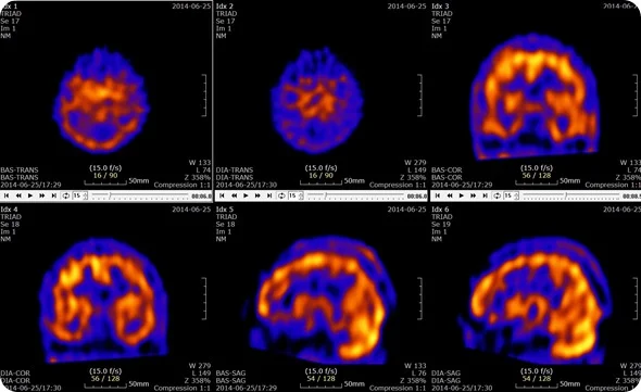
A SPECT scan provides 3-D images of the distribution of radioactive tracer molecules in the patient’s body. The images are computer generated from projection images of the body that are recorded at different angles by the SPECT machine. SPECT machines have gamma radiation detectors that can detect the gamma-ray radiation emitted from the tracers injected into the patient.
You can see how the scan is performed here.
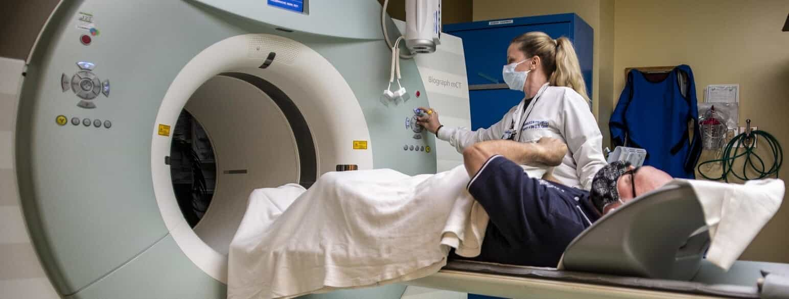
What is a PET scan?
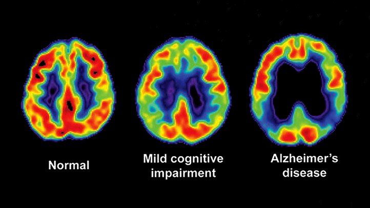
A PET scan can help reveal your tissues and organs’ metabolic or biochemical function. Like with SPECT, PET scans use radioactive tracers to show typical and atypical metabolic activity. Cancer cells have a higher metabolic activity than normal cells, which makes PET scans useful for cancer detection. PET scans can also be used to detect heart disease and brain disorders like Alzheimer’s and dementia.
You can see how a PET scan is performed here.
Why is nuclear medicine helpful?
SPECT scans can detect and monitor the progression of heart disease, such as blocked coronary arteries. Coronary arteries supply blood to the heart muscle, and a lack of blood flow can lead to ischemic heart disease, which can cause heart attacks and death. Other radiotracers can detect bone diseases, gallbladder disease, and intestinal bleeding. SPECT has also been used to help diagnose Parkinson’s disease in the brain and distinguish it from other anatomically comparable movement disorders and dementias. As mentioned earlier, PET scans play an important role in cancer detection and heart disease monitoring.
What are the risks of using radioactive tracers?
Given its name, nuclear medicine exposes the patient to radiation. This dose is no more than what is experienced during routine chest X-ray or CT exams. As a result of radiation exposure, these procedures increase your risk of developing cancer later in life. However, this risk is considered to be minimal compared to the expected benefit from a medically necessary diagnostic imaging exam. Physicians agree that the pros of undergoing nuclear medicine examinations outweigh the cons of not undergoing these procedures. With imaging, some conditions may be noticed, or confirmation of diagnosis cannot be made, which would lead to better outcomes for the patient.
The nuclear medicine team, which consists of nuclear medicine technicians, medical physicists, and nuclear medicine-specialized physicians, is committed to tailoring the dose of radiation needed for each patient so that it’s the lowest effective dose. Due to these specialists, nuclear medicine imaging is very safe. As a result, only a few people are contraindicated from undergoing nuclear medicine procedures. The most common contraindications include pregnancy (to avoid exposure of the fetus to radiation) or allergic reactions to the radioactive tracers.
Conclusion
Nuclear medicine is a vital field that uses radioactive tracers for detailed diagnostic imaging, providing insights into the health and function of organs and tissues. Despite common associations with nuclear weapons and power plants, in medicine, “nuclear” refers to a safe and powerful tool for diagnosing conditions such as, but not limited to, heart disease, bone disorders, Parkinson’s disease, and various cancers. Utilizing SPECT and PET scans, nuclear medicine offers unparalleled accuracy in detecting and monitoring these diseases. While radiation exposure poses minimal risk, the benefits of precise diagnosis and effective treatment planning far outweigh this concern. With careful dose management by specialized professionals, nuclear medicine continues to be an indispensable component of modern healthcare, driving better patient outcomes and advancing our ability to combat complex diseases.
References
https://www.nibib.nih.gov/science-education/science-topics/nuclear-medicine
https://www.hopkinsmedicine.org/health/treatment-tests-and-therapies/nuclear-medicine
https://www.cdc.gov/radiation-health/data-research/facts-stats/nuclear-medicine.html
Image credits:
Cover Photo: AI
Figure 1. https://live.staticflickr.com/1826/42436453085_62fe1c6c00_b.jpg
Figure 2. https://media.lex.dk/media/179425/article_topimage_PET-scanning.jpg

[…] Arbona-Lampaya explains nuclear medicine and its significance (p. […]
LikeLike